Ultrasound/sonograms are sound waves with frequencies higher than the upper audible limit of human hearing. In the medical field, ultrasound imaging or diagnostic sonography is a medical test that uses sound waves or echoes to capture and produce live images of internal body structures. The ultrasonic images produced are also called sonograms. Ultrasound is based on sonar and uses a machine with a computer processor to create Ultrasound images.

These sonograms are used in clinics and hospital settings by doctors and other medics to help diagnose the causes of pain, swelling, and
infection in the body's internal organs and to examine a baby in pregnant women and the brain and hips in infants. They also assist them in
guiding biopsies, diagnose heart conditions, and assess damage after a heart attack. Ultrasound is safe, non-invasive, and does not use
ionizing radiation.
Ultrasound waves are produced by a transducer made of crystal materials that can both emit ultrasound waves, as well as detect the
ultrasound echoes reflected back. When used in an ultrasound scanner, the transducer sends out a beam of sound waves into the body. The
sound waves are reflected back to the transducer by boundaries between tissues in the path of the beam (e.g. the boundary between fluid and
soft tissue or tissue and bone). When echoes hit the transducer, they generate electrical
signals that are sent to the ultrasound scanner.
Using the speed of sound and the time of each echo’s return, the scanner calculates the distance from the transducer to the tissue boundary.
These distances are then used to generate two-dimensional images of tissues and organs.
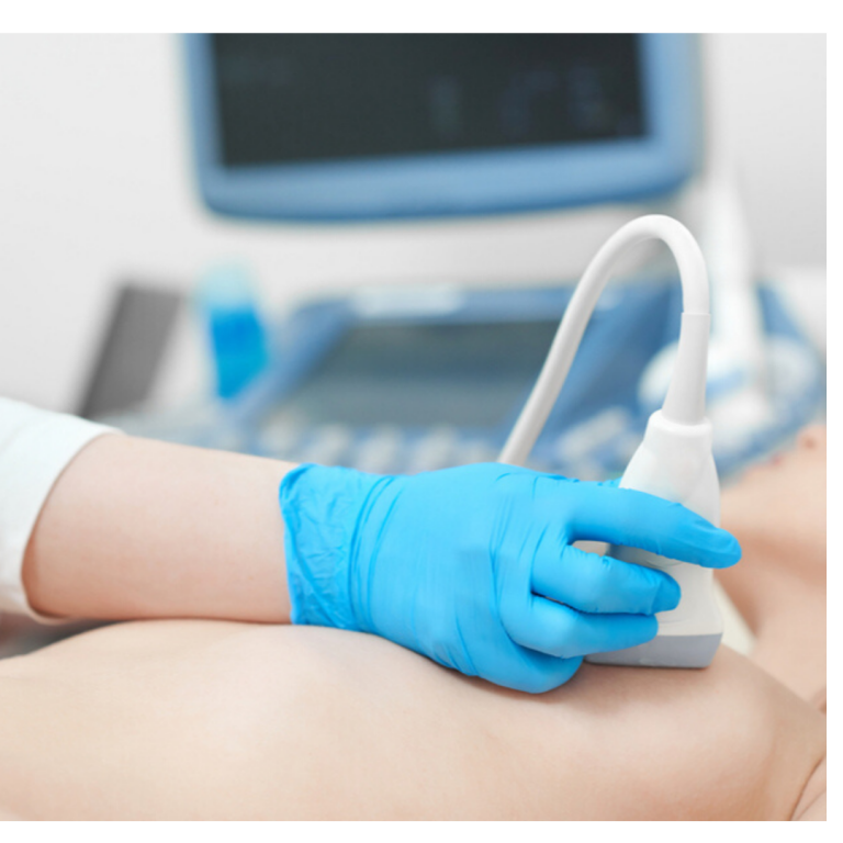
During an ultrasound exam, the technician will apply a gel to the skin. This keeps air
pockets from forming between the transducer and the skin, which can block
ultrasound waves from passing into the body.
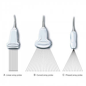
Diagnostic ultrasound is a non-invasive diagnostic technique that uses ultrasound probes, also called transducers to image inside the body. The transducers are placed on the skin or inside the body through the vagina, the gastrointestinal tract or blood vessels. Diagnostic ultrasound. Diagnostic ultrasound is able to non-invasively image internal organs within the body. However, it is not good for imaging bones or any tissues that contain air, like the lungs. Under some conditions, ultrasound can image bones (such as in a fetus or in small babies) or the lungs and lining around the lungs, when they are filled or partially filled with fluid. One of the most common uses of ultrasound is during pregnancy, to monitor the growth and development of the fetus, but there are many other uses, including imaging the heart, blood vessels, eyes, thyroid, brain, breast, abdominal organs, skin, and muscles. Ultrasound images are displayed in either 2D, 3D, or 4D (which is 3D in motion).
The ultrasound probe (transducer) is placed over the carotid artery (top). A color ultrasound image (bottom, left) shows blood flow (the red
color in the image) in the carotid artery. Waveform image (bottom right) shows the sound of flowing blood in the carotid artery.
Functional ultrasound. Functional ultrasound applications include Doppler and color Doppler ultrasound for measuring and visualizing
blood flow in vessels within the body or in the heart. It can also measure the speed of the blood flow and direction of movement. This is done
using color-coded maps called color Doppler imaging. Doppler ultrasound is commonly used to determine whether plaque build-up inside
the carotid arteries is blocking blood flow to the brain.
Another functional form of ultrasound is elastography, a method for measuring and displaying the relative stiffness of tissues, which can
be used to differentiate tumors from healthy tissue. The images can be displayed as either color-coded maps of the relative stiffness; black-
and white maps that display high-contrast images of tumors compared with anatomical images; or color-coded maps that are overlayed on
the anatomical image. Elastography can be used to test for liver fibrosis, a condition in which excessive scar tissue builds up in the liver due
to inflammation.
Therapeutic or interventional ultrasound. This produces high levels of acoustic output that can be focused on specific targets for the
purpose of heating, ablating or breaking up tissue. Therapeutic ultrasound produces high levels of acoustic output that can be focused on
specific targets for the purpose of heating, ablating, or breaking up tissue. also uses sound waves above the range of human hearing but does
not produce images. Its purpose is to interact with tissues in the body such that they are either modified or destroyed. One good thing about
this ultrasound is that no incisions or cuts need to be made to the skin, leaving no wounds or scars.
One type of therapeutic ultrasound uses high-intensity beams of sound that are highly targeted, and is called High Intensity Focused
Ultrasound (HIFU). HIFU is being investigated as a method for modifying or destroying diseased or abnormal tissues inside the body (e.g.
tumors) without having to open or tear the skin or cause damage to the surrounding tissue. Either ultrasound or MRI is used to identify and
target the tissue to be treated, guide and control the treatment in real time, and confirm the effectiveness of the treatment.
Ultrasound is also an important method for imaging interventions in the body. For example, ultrasound-guided needle biopsy helps
physicians see the position of a needle while it is being guided to a selected target, such as a mass or a tumor in the breast. Also, ultrasound is
used for real-time imaging of the location of the tip of a catheter as it is inserted in a blood vessel and guided along the length of the vessel. It
can also be used for minimally invasive surgery to guide the surgeon with real-time images of the inside of the body.
Abdominal Ultrasound
An abdominal ultrasound is a useful way of examining internal organs, including the liver, gallbladder, spleen, pancreas, kidneys, and bladder. This can help to diagnose a variety of conditions and to assess the damage caused by illness.
Pelvic ultrasound
Pelvic ultrasound is often used to diagnose conditions or the cause of conditions such as: Pelvic pain, Abnormal bleeding, Menstrual
problems, Ovarian cysts, Uterine fibroids, Ovarian and uterine cancers, Kidney and bladder stones, Transabdominal. Patients receiving a
transabdominal ultrasound need to have a full urinary bladder. The transducer is then rubbed over the examination are and releases sound waves.
Transvaginal Ultrasound
During this procedure, a woman needs to empty her bladder the same way she would for a gynecological exam. She also lies face up on her
back with feet in stirrups. The transducer of the ultrasound needs to be inserted for this test. The transducer is smaller than the standard
speculum used in Pap tests. A protective cover and gel for lubrication is placed on the transducer before it is inserted in the vagina.
Only the first two to three inches of the transducer is inserted in the vagina. The doctor may move it around to obtain images from different
angles. The most common reason for transvaginal pelvic ultrasounds is to look for the cause of pelvic pain.
Transrectal
In order to perform an ultrasound on the prostate gland, the transducer must be inserted through the rectum so that the sound waves can
travel to the prostate. As with other inserted ultrasound procedures, the transducer is covered with a protective cover and lubrication before
insertion. A transducer will need to be moved around in order to obtain images from different angles.
Obstetric Ultrasound Imaging
Obstetric ultrasound (OB ultrasound) refers to the specialized use of sound waves to visualize and thus determine the condition of a pregnant
woman and her embryo or fetus. Obstetric ultrasound should be performed only when clinically indicated. Some indications may be to
establish the presence of a living embryo/fetus, to estimate the age of the pregnancy, to diagnose congenital abnormalities, to evaluate the
position of the fetus or the position of the placenta and to determine if there are multiple pregnancies.
This is a painless procedure. There may be varying degrees of discomfort from pressure as the sonographer guides the transducer over your
abdomen, especially if you are required to have a full bladder. At times the sonographer may have to press more firmly to get closer to the
embryo or fetus to better visualize the structure. This discomfort is temporary. Also, you may dislike the feeling of the water-soluble gel
applied to your abdomen. With transvaginal scanning, there may be minimal discomfort as the transducer is moved in the vagina.
Please note that we do not permit video recordings or photos from any recording devices (i.e. cell phone, camera, etc.) in the exam room. In
addition, our ultrasound rooms are not large and we may need to limit the number of guests during your scan. There are many aspects of the
pregnancy that the sonographer needs to assess during your ultrasound and therefore may limit discussion during the exam.
Carotid & Abdominal Aorta Ultrasound Imaging
Ultrasound of the carotid arterial system provides a fast, noninvasive means of identifying blockages of blood flow in the neck arteries to the
brain that might produce a stroke or mini-stroke. Ultrasound of the abdominal aorta is primarily used to evaluate for an aneurysm which is
an abnormal enlargement of the aorta usually from atherosclerotic disease.
The patient is positioned on an examination table that can tilt and move. A clear gel is applied to the area that will be examined. The gel helps
the transducer make a secure contact and eliminates air pockets between the transducer and the skin, since the sound waves cannot
penetrate air. The sonographer or radiologist then presses the transducer firmly against the skin and sweeps along the area of interest,
reviewing the images on the monitor and capturing "snapshots" as required.
Liver Ultrasound
Liver Ultrasound determines size, shape, and function of the liver, and can be used to detect tumors.
Renal Ultrasound
It determines the size, shape, and function of the kidneys, and can be useful in the detection of kidney stones, cysts, and tumors.
Vascular Ultrasound
Vascular ultrasounds are used to analyze the flow of blood through the arteries and veins.
Thyroid Ultrasound
Thyroid Ultrasound checks for underactive and overactive thyroid glands, nodules, and cysts.
A Doppler ultrasound is a special ultrasound technique that evaluates the movement of materials in the body. It allows the doctor to see and evaluate blood flow through arteries and veins in the body.
Color Doppler uses a computer to convert Doppler measurements into an array of colors to show the speed and direction of blood flow
through a blood vessel.
Power Doppler is a newer technique that is more sensitive than color Doppler and capable of providing greater detail of blood flow, especially
when blood flow is little or minimal. Power Doppler, however, does not help the radiologist determine the direction of blood flow, which may
be important in some situations.
Spectral Doppler displays blood flow measurements graphically, in terms of the distance traveled per unit of time, rather than as a color
picture. Also converts blood flow information into a distinctive sound that can be heard with every heartbeat.
Doppler ultrasound images can help the physician to see and evaluate:
Blockages to blood flow (such as clots)
Narrowing of vessels
Tumors and congenital vascular malformations
Reduced or absent blood flow to various organs, such as the testes or ovary
Increased blood flow, which may be a sign of infection
Benefits of Ultrasound Machines
Most ultrasound scanning is noninvasive (no needles or injections).
Occasionally, an ultrasound exam may be temporarily uncomfortable, but it should not be painful.
Widely available, easy-to-use and less expensive than most other imaging methods.
Extremely safe and does not use radiation.
Gives a clear picture of soft tissues that do not show up well on x-ray images.
Preferred imaging modality for the diagnosis and monitoring of pregnant women and their unborn babies.
Provides real-time imaging, making it a good tool for guiding minimally invasive procedures like biopsies.
https://www.nibib.nih.gov/science-education/science-topics/ultrasound
https://www.radiologyinfo.org/en/info.cfm?pg=genus
https://en.wikipedia.org/wiki/Medical_ultrasound
For more information, you can contact us via email or give us a call.
Please share your thoughts on the comments section below.