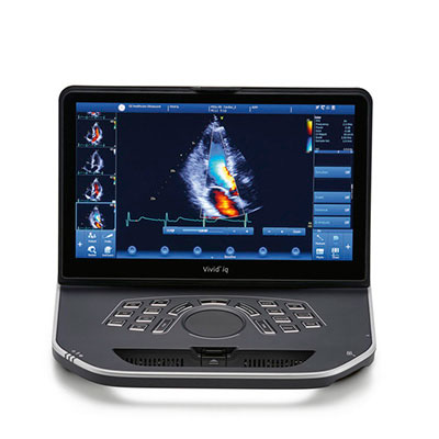
The GE Vivid IQ is a portable ultrasound system with an integrated full touch screen control, conventional user control panel, and trackpad design instead of the trackball interface.
The design of the GE Vivid IQ is lightweight, moreover, come with one DLP and three RS to support 6VT-D ports and a dedicated cart.
The GE Vivid IQ ultrasound machine enables to support of musculoskeletal, adult cephalic, neonatal cephalic, small organ, pediatric, abdominal adults, fetal/OB, peripheral vascular, transesophageal, and cardiac ultrasounds.
GE Vivid IQ Dimensions and Weight
GE Vivid IQ Specifications
GE Vivid IQ Electrical power
Review
The second generation of GE Vivid IQ is an all-new system that has been developed based on Vivid I and Q.
The ultrasound machine has an experimental interface and hardware feature.
The console design is completely different from the former Vivid Q and I system, integrated with a multi-touch screen of 15.6 that enables users to experience a more intuitive workflow.
The ultrasound machine uses a trackpad instead of a trackball that is currently being used by most systems.
Auto-Optimization
The images can be optimized with the one-touch image optimization on the actual pulse wave doppler data and B-mode image.
For contrast enhancement in the resulting image, the function allows users to pick preference and works best on preset levels, low, medium.
Low dose, compared to high dose, does the least amount of contrast enhancement, whereas, the high dose does the most.
Auto-optimization is present in Spectral Doppler, while in zoom, CINE (in B-mode only), frozen, live images in a multi or single image.
PW Doppler Mode uses auto-optimization to optimize the spectral data. The invert (if preset), dynamic range, baseline shift, and velocity scale can be adjusted with auto-adjusts. The spectrum is optimized even upon deactivation.
B-Flow
B-Flow visualizes blood flow according to hemodynamics and reflectors speed using grayscale imaging with different gray intensities. B-Flow does not depend B-Flow may help in visualizing bladder reflux or jets, liver and spleen vasculature, kidney perfusion, and vessel wall irregularities.
Anatomical M-Mode
The Anatomical M-Mode helps improve cardiac measurements and arrhythmia assessment accuracy. It enables M-Mode images perpendicular to the ventricular septum.
Raw Data
The software tool enables image analysis, quick data re-acquisition, and image processing. Moreover, reduces time to put the probe on patient, improves clinical workflow, and shortens exam duration.
Auto IMT
Auto IMT automates the measurement of intima-media thickness of vessels and helps keep track of atherosclerosis disease as it is developed, from the early stage.
Auto EF
Auto EF automatically measures LV ejection fraction, maximizing time efficiency.