In medicine, endoscopy is a non-surgical procedure that involves the insertion of a long, thin tube directly into the body to observe an internal organ or tissue in detail.
An endoscope is an illuminated optical, typically slender and tubular instrument used to look deep into the body with a light and camera attached to it.
In 1806, the first endoscope was designed then later developed at Glasgow Royal Infirmary in Scotland in 1894/5 by Dr John Macintyre as part of his specialization in the investigation of the larynx.
When you visit the hospital, your doctor can view pictures of your internal organs on a color TV monitor.
Endoscopy is a minimally invasive process used for procedures such as imaging and minor surgeries. Although painless, it can be pretty uncomfortable at times.
I mean, having a tube moving down your throat all the way to your lungs or up your rectum is not funny as it sounds, nope. Well, endoscopy can either be upper or lower. We shall look into that shortly.
One of the upper procedures is known as bronchoscopy. A bronchoscope is used by the doctor to examine the airways and diagnose lung disease.
Lower endoscopy, on the other hand, allows your doctor to pass the endoscope through the anus into the large intestine/colon, through the rectum for examination.
This procedure is called sigmoidoscopy or colonoscopy depending on how far up the colon is examined. Alternatively, the endoscope can be inserted for small incisions like the urethra, skull, knee, or chest.
We will be digging deeper into the processes carried out during endoscopy procedures so that your next visit to the doctor may be a walk in the park. Ready? Now, let's get into it. √
We have hundreds of tissues in our body and so are the number of procedures to examine them. Today, we'll not talk about them all, just a chosen few. Earlier we mentioned about the two classes of endoscopy; upper and lower. Let's break them down.

Endoscopy procedure
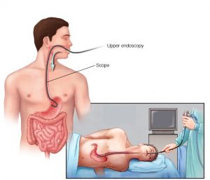
An upper endoscopy is also called an upper GI endoscopy or esophagogastroduodenoscopy (EGD). It allows the medical personnel to look at the upper part of the gastrointestinal (GI) tract.
This area is made up of the: esophagus, which is the muscular tube that connects the throat to the stomach, stomach, and duodenum, which is the top of the small intestine.
Doctors carrying out upper endoscopy, also known as gastroenterologists or GI specialists, usually pass the endoscope through the mouth and throat into the esophagus, and finally to the upper part of the small intestine/duodenum. This allows the doctor to view these organs on a color TV.
A special form of endoscopy called endoscopic retrograde cholangiopancreatography, or ERCP, allows pictures of the pancreas, gallbladder, and related structures to be taken.
ERCP is also used for stent placement and biopsies. Endoscopic ultrasound or EUS combines upper endoscopy and ultrasound examination to obtain images and information about various parts of the digestive tract.
Doctors will often recommend endoscopy to evaluate:
Stomach pain
Ulcers, gastritis, or difficulty swallowing
Digestive tract bleeding
Changes in bowel habits (chronic constipation or diarrhea)
Polyps or growths in the colon
In addition, your doctor may use an endoscope to take a biopsy (removal of tissue) to look for the presence of disease.
Before the procedure begins, you will need to undress and put on a hospital gown. If you wear dentures, you may be asked to remove them.
You may be given anesthesia and a sedative through an intravenous (IV) needle in your arm. Anesthesia is a medicine that blocks the awareness of pain. A sedative is a medicine that relaxes you. These medications help lessen discomfort during the procedure. You may feel a slight sting when the IV needle is inserted.
A doctor or nurse may spray your throat with a local anesthetic to numb it. Or you may receive a liquid anesthetic to gargle. This helps prevent gagging. The doctor may also insert a mouthpiece to hold your mouth open during the procedure.
You will lie on your left side or your back on an exam table. As the doctor slowly inserts the endoscope, you will feel pressure through your esophagus. The endoscope inflates your stomach by blowing air into it. This gives the doctor a better view of the stomach lining.
You will be able to breathe on your own throughout the exam. Taking slow, deep breaths through your nose will help you relax. Many people fall asleep.
An upper endoscopy usually takes 20 to 30 minutes to complete. When the procedure is over, the doctor will gently remove the endoscope. Then you will go to a recovery room.
Endoscopy may also be used to treat a digestive tract problem. For example, the endoscope might not only detect active bleeding from an ulcer but devices can be passed through the endoscope that can stop the bleeding. In the colon, polyps can be removed through the scope to prevent the development of colon cancer.
It may be used to investigate symptoms in the digestive system including nausea, vomiting, abdominal pain, difficulty swallowing, and gastrointestinal bleeding.
It is also used in diagnosis, most commonly by performing a biopsy to check for conditions such as anemia, bleeding, inflammation, and cancers of the digestive system.
The procedure may also be used for treatment such as cauterization of a bleeding vessel, widening a narrow esophagus, clipping off a polyp, or removing a foreign object.
Lower gastrointestinal (GI) endoscopy is a minimally invasive diagnostic technique that allows the clinician to evaluate the mucosal surfaces of the rectum, colon, ileocolic sphincter, cecum, and distal small intestine (ileum).
The procedure may be utilized to Screen for cancers, investigate symptoms, diagnose gastrointestinal diseases and disorders, control bleeding, extract polyps, foreign objects, tissue samples for biopsy, among others.
Colonoscopy. Also known as coloscopy, this is the endoscopic examination of the large bowel and the distal part of the small bowel with a CCD camera, a fiber optic camera on a flexible tube passed through the anus.
Proctoscopy. This is a common medical procedure in which an instrument called a proctoscope is used to examine the anal cavity, rectum, or sigmoid colon.
Ileoscopy. It is an endoscopic procedure that uses a tiny camera attached to a scope to examine the lower portion of the small intestine (the ileum).
Colonoscopy can take 30 minutes or longer. Sigmoidoscopy often takes about 20 minutes. The length of the procedure depends a great deal on how clean your intestines are, the reason for the procedure, and what treatments must be done.
You lie on the stretcher or bed on your left side.
For colonoscopy, you are given sedating (relaxing) medicine through an IV line. Sigmoidoscopy usually doesn’t need sedation.
The endoscope is inserted into your rectum. You may feel pressure and cramping. If you feel pain, tell your healthcare provider. You may receive more sedation, which includes pain medicine and an anti-anxiety medicine.
The endoscope carries images of your colon to a video screen. Prints of the images may be taken as a record of your exam.
Biopsies (tissue samples), polyp removal, or other treatments may be performed.
When the procedure is done, you rest for a time. You may have some discomfort right after the procedure from trapped air. This can be relieved by changing position and passing the air. If you have been sedated, you must have an adult drive you home.
Type of Tools Used in Endoscopy;
Flexible forceps. These tong-like tools take a tissue sample.
Biopsy forceps. These remove a tissue sample or a suspicious growth.
Cytology brushes. These take cell samples.
Suture removal forceps. These remove stitches inside the body
An anoscope, a proctoscope, and a rectoscope with approximate lengths
Endoscopy surgery
Health care providers can use endoscopy to review any of the following body parts:
The gastrointestinal tract (GI tract):
Esophageal Bougie Dilator, esophagus, stomach and duodenum (esophagogastroduodenoscopy), small intestine (enteroscopy)
large intestine/colon (colonoscopy, sigmoidoscopy).
Magnification endoscopy, bile duct, endoscopic retrograde cholangiopancreatography (ERCP), duodenoscope-assisted cholangiopancreatoscopy, intraoperative cholangioscopy, rectum (rectoscopy) and anus (anoscopy), both also referred to as (proctoscopy).
The respiratory tract, The nose (rhinoscopy), The upper respiratory tract (laryngoscopy), The lower respiratory tract (bronchoscopy).
The ear (otoscope), The urinary tract (cystoscopy), The female reproductive system (gynoscopy), The cervix (colposcopy).
The uterus (hysteroscopy), The fallopian tubes (falloposcopy), Normally closed body cavities (through a small incision):
The abdominal or pelvic cavity (laparoscopy), The interior of a joint (arthroscopy), Organs of the chest (thoracoscopy and mediastinoscopy)
Endoscopy is used for many procedures:
During pregnancy
The amnion (amnioscopy), The fetus (fetoscopy), Plastic surgery, Panendoscopy (or triple endoscopy), Combines laryngoscopy, esophagoscopy, and bronchoscopy, Orthopedic surgery, Hand surgery, such as endoscopic carpal tunnel release, Knee surgery, such as anterior cruciate ligament reconstruction, Epidural space (Epiduroscopy), Bursae (Bursectomy), Endodontic surgery, Maxillary sinus surgery, Apicoectomy, Endoscopic endonasal surgery, Endoscopic spinal surgery.
An Endoscopy is a simple procedure which allows a doctor to look inside human bodies using an instrument called an endoscope. A cutting tool can be attached to the end of the endoscope, and the apparatus can then be used to perform surgery. This type of surgery is called Key hole surgery, and usually leaves only a tiny scar externally.
Capsule endoscopy was developed in the mid-1990s and involves a wireless camera. The camera is small enough to fit into a capsule (roughly the size of a vitamin tablet) and can, therefore, be swallowed. As the capsule travels through the digestive tract, it takes thousands of pictures, which are transmitted to a device attached to a wearable belt. This procedure is used to image the small intestine, a region that is difficult to image using standard endoscopy. It is also very useful for examining the small intestinal mucosa and diagnosing Crohn’s disease. The capsule usually passes through the digestive system within 24-48 hours.
Perforation (tear in the gut wall)
Reaction to sedation
Infection
Bleeding
Pancreatitis as a result of ERCP
https://www.medicalnewstoday.com/articles/153737
https://www.webmd.com/digestive-disorders/digestive-diseases-endoscopy#1
https://www.mayoclinic.org/tests-procedures/endoscopy/about/pac-20395197
https://en.wikipedia.org/wiki/Endoscopy
Share with us your past experiences or upcoming expectations on endoscopy procedures.
You must be logged in to post a comment.

nice article
thank you